Tendon Diagram / Cunningham S Text Book Of Anatomy Anatomy 346 The Akticulations Oe Joints Forwards Subjacent To This Ligament. The tendon sheath of the posterior tibial muscle covers the posterior and middle part of the deltoid ligament in much the same way as the peroneal tendon sheath is associated with the calcaneofibular ligament on the lateral side. Raises heal when leg is straight. Fall on one point of shoulder and can rupture these ligaments with dislocation of ac joint. Ligaments join the knee bones and provide stability to the knee: On the other hand, the insertion is where a tendon attaches that muscle to the *more* movable bone.
In human anatomy, a hamstring (/ ˈ h æ m s t r ɪ ŋ /) is any one of the three posterior thigh muscles in between the hip and the knee (from medial to lateral: Also allows the action of raising up onto toes. The hamstrings are quite susceptible to injury. The tendon sheath of the posterior tibial muscle covers the posterior and middle part of the deltoid ligament in much the same way as the peroneal tendon sheath is associated with the calcaneofibular ligament on the lateral side. A tendon is a band of tissue that connects a muscle to a bone.
Diagram Right Arm Tendon Diagram Full Version Hd Quality Tendon Diagram Agenciadiagrama I Ras It from 1.bp.blogspot.com The hamstrings are quite susceptible to injury. Hand a hand is a prehensile multi fingered appendage located at the end of the forearm or forelimb of primates such as humans chimpanzees monkeys and lemurs human anatomy for the artist the dorsal hand the dorsal the easiest tendons to identify in the dorsal hand are those of the extensor digitorum muscle its name means extensor of the digits which is Brings hip away from body. Both are made of collagen.ligaments connect one bone to another, while tendons connect muscle to bone. A muscle's origin is where a tendon attaches it to the *less* movable bone. A tendon is a band of tissue that connects a muscle to a bone. The hip itself is a ball and socket joint, much like the shoulder.the structures necessary to create this joint are the socket, the joint capsule, muscle, ligaments, and the neck. Tendon diagram simple / 8.4c:
Brings hip away from body.
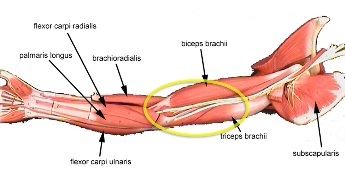
Movement occurs when our muscles pull on our bones, relocating them. The changes in ligaments and tendons generally occur more slowly than adaptation in bone, because ligaments and tendons have less vascular supply. The shoulder joint is formed the rotator cuff is a collection of muscles and tendons that. Tendons, located at each end of a muscle, attach muscle to bone. Tendons are thick bands of tissue that connect muscles to bones. A muscle's origin is where a tendon attaches it to the *less* movable bone. Tendons are found throughout the body, from the head and neck all the way down to the feet. In the back and elsewhere in the body, tendons attach muscles to bones. Flexes elbow and moves forearm. This important tendon in the back of the calf and ankle connects the plantaris, gastrocnemius, and soleus muscles to. Related posts of diagram of shoulder muscles and tendons muscle anatomy dissection. Your biceps tendons attach the biceps muscle to bones in the shoulder and in the elbow. Also allows the action of raising up onto toes.
Diagram showing the tendons and ligaments of the ankle and. Again, our knowledge of how mechanical stimulus mediates ligament and tendon structure is more empirical and less. 2 ligaments (trapezoid& conoid ligaments) attach the clavicle coracoid process of scapula these tiny ligaments (w/ acominoclavicular joint) keep scapula attached to clavicle. A tendon is a tough yet flexible band of fibrous tissue. Posted on april 3, 2019april 3, 2019.
Body Anatomy Upper Extremity Tendons The Hand Society from www.assh.org Learn about the anatomy and physiology of tendons. The bones together make up the hip. The achilles tendon is also called the calcaneal tendon. Fall on one point of shoulder and can rupture these ligaments with dislocation of ac joint. Diagram showing the tendons and ligaments of the ankle and. The achilles tendon is the largest. One peroneal tendon attaches to the outer part of the midfoot, while the other tendon runs under the foot and attaches near the inside of the arch. This important tendon in the back of the calf and ankle connects the plantaris, gastrocnemius, and soleus muscles to.
In human anatomy, a hamstring (/ ˈ h æ m s t r ɪ ŋ /) is any one of the three posterior thigh muscles in between the hip and the knee (from medial to lateral:
Biceps and triceps tendon rupture. The tendon connects muscle to the bone. Tendons are thick bands of tissue that connect muscles to bones. Tendon diagram simple / 8.4c: Ligaments and tendons are adapted in response to changes in mechanical stiffness. Its muscle belly is in the forearm. In human anatomy, a hamstring (/ ˈ h æ m s t r ɪ ŋ /) is any one of the three posterior thigh muscles in between the hip and the knee (from medial to lateral: Numerous muscles help stabilize the three joints of. Ligaments join the knee bones and provide stability to the knee: Brings hip away from body. Tendon, tissue that attaches a muscle to other body parts, usually bones. The achilles tendon is the largest. Diagram depicting the bones, ligaments and muscles throughout the hand and fingers.
Raises and rotates arm in all directions. If you tear the biceps tendon at the shoulder, you may lose some strength in your arm and have pain when you forcefully turn your arm from palm down to palm up. The tendon sheath of the posterior tibial muscle covers the posterior and middle part of the deltoid ligament in much the same way as the peroneal tendon sheath is associated with the calcaneofibular ligament on the lateral side. 2 ligaments (trapezoid& conoid ligaments) attach the clavicle coracoid process of scapula these tiny ligaments (w/ acominoclavicular joint) keep scapula attached to clavicle. The anterior cruciate ligament prevents the femur from sliding backward on the tibia (or the tibia sliding forward on the femur).
Flexor Tendon Injuries Fife Virtual Hand Clinic from fifevirtualhandclinic.files.wordpress.com Elbow muscle anatomy mri 12 photos of the elbow muscle anatomy mri elbow muscle anatomy axial, elbow muscle anatomy mri, human muscles, elbow muscle anatomy axial, elbow muscle anatomy mri The two peroneal tendons in the foot run side by side behind the outer ankle bone. Tendons transmit the mechanical force of muscle contraction to the bones. Tendon, tissue that attaches a muscle to other body parts, usually bones. The bones together make up the hip. It is constructed in such a way that we can move the arms to. Tendons, located at each end of a muscle, attach muscle to bone. Related posts of muscles and tendons of the leg elbow muscle anatomy mri.
Also allows the action of raising up onto toes.
Your biceps tendons attach the biceps muscle to bones in the shoulder and in the elbow. Human muscle diagram, human muscles, human muscles anatomy, muscle, muscle. Raises and rotates arm in all directions. 2 ligaments (trapezoid& conoid ligaments) attach the clavicle coracoid process of scapula these tiny ligaments (w/ acominoclavicular joint) keep scapula attached to clavicle. The tendon connects muscle to the bone. Flexes elbow and moves forearm. Muscles of the shoulder : Biceps and triceps tendon rupture. The achilles tendon is also called the calcaneal tendon. Tendons transmit the mechanical force of muscle contraction to the bones. A muscle's origin is where a tendon attaches it to the *less* movable bone. Brings leg back to and across body. Both are made of collagen.ligaments connect one bone to another, while tendons connect muscle to bone.